August 6, 2021
Overview
Overview
- A brief introduction of segmentation analysis and ChromHMM
- Apply the analysis to a test dataset and explore the output
- Interpret the output of the segmentation using functions from
segmenter
Background
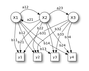
Hidden Markov Models (HMM)
ChromHMM
- ChromHmm is a Java program to learn chromatin states from multiple sets of histone markers ChIP-seq data
- States are modeled as the combination of markers on the different regions of the genome.
- A multi-variate hidden Markov model is used to model the presence or absence of the markers.
- The combination of histone markers at a given location is interpreted as a biological state.
- Ref: Ernst, J., Kellis, M. Chromatin-state discovery and genome annotation with ChromHMM. Nat Protoc 12, 2478–2492 (2017).
This package!
The goal of the segmenter package is to
- Call ChromHMM using R syntax
- Capture the output in R objects
- Interact with the model output for the purposes of summarizing or visualizing
Getting Started
Installation
The package can be installed from Bioconductor using BiocManager or from GitHub using remotes
# install from bioconductor
BiocManager::install('segmenter')

# install from github
remotes::install_github('MahShaaban/segmenter@devel')
Example code
# run command
obj <- learn_model(
    inputdir = <input binarized files>,
    coordsdir = <genomic coordinates of features>,
    anchorsdir = <genomic location of TSS>,
    chromsizefile = <chromosome lengths>,
    numstates = <the number of desired states>,
    assembly = <the genome assembly>,
    cells = <names of the biological samples>,
    annotation = <name of the reference genome type>,
    binsize = <the size of bins>
)
Using segmenter
Inputs
ChromHMM requires two types of input files. Those are
- Genomic annotation files
- Coordinates: the start and end location of genomic features to calculate enrichment
- Anchors: the transcription start and end sites
- Chromosome size: the length of each chromosome
- Binarized signal files from the ChIP-seq data
Genomic annotation files
# coordinates
coordsdir <- 'hg38_coords'
list.files(coordsdir)
#> [1] "CpGIsland.hg38.bed.gz"    "RefSeqExon.hg38.bed.gz"
#> [3] "RefSeqGene.hg38.bed.gz"   "RefSeqTES.hg38.bed.gz"
#> [5] "RefSeqTSS.hg38.bed.gz"    "RefSeqTSS2kb.hg38.bed.gz"

# anchors
anchorsdir <- 'hg38_anchors'
list.files(anchorsdir)
#> [1] "RefSeqTES.hg38.txt.gz" "RefSeqTSS.hg38.txt.gz"

# chromosomes' sizes
chromsizefile <- 'hg38_chromsizes.txt'
read.delim(chromsizefile, header = FALSE, nrows = 2)
#>     V1        V2
#> 1 chr1 248956422
#> 2 chr2 242193529
Binarized signal files from the ChIP-seq data
- The binarized signal files are text files, often one for each chromosome
- Data is divided into bins of a given size (rows) and have binary values 1 (> threshold) or 0 for each histone markers (columns)
- From bam
binarize_bamor bedbinarize_bed
# a table to assign marker and cell names to the bam files
cell_marks <- 'cell_mark_file.tsv'
read.delim(cell_marks, header = FALSE, nrows = 3)
#>        V1       V2              V3
#> 1 GM12878 H3K27me3 ENCFF796DDM.bam
#> 2 GM12878 H3K27me3 ENCFF927XRX.bam
#> 3 GM12878 H3K27me3 ENCFF265UBT.bam
# run command
binarize_bam('bam/',
             chromsizefile = chromsizefile,
             cell_marks = cell_marks,
             outputdir = 'bins_200')
# show output files
example_binaries <- list.files('bins_200', full.names = TRUE)[1]
read.delim(example_binaries, nrows = 2, skip = 1)
#>   H2AFZ H3K27ac H3K27me3 H3K36me3 H3K4me1 H3K4me2 H3K4me3 H3K79me2 H3K9ac
#> 1     0       0        0        0       0       0       0        0      0
#> 2     0       0        0        0       0       0       0        0      0
#>   H3K9me3 H4K20me1
#> 1       0        0
#> 2       0        0
Model Learning
learn_model wraps the the Java module that learns a chromatin segmentation model of a given number of states.
# load library
library(segmenter)

# run the main command
obj <- learn_model(
    inputdir = 'bins_200',
    outputdir = 'models/model_10',
    anchorsdir = 'hg38_anchors/',
    coordsdir = 'hg38_coords/',
    chromsizefile = 'hg38_chromsizes.txt',
    numstates = 10,
    cells = c('GM12878', 'H1', 'HepG2', 'IMR-90', 'K562'),
    assembly = 'hg38',
    annotation = 'RefSeq',
    binsize = 200
)
Output
# show the object
class(obj)
#> [1] "segmentation"
#> attr(,"package")
#> [1] "segmenter"
model: the initial and final parametersemission: the probabilities of the mark in a given statetransition: the probabilities of the states transitionsoverlap: the enrichment of the states at genomic featuresTSS: the enrichment of the states around the TSSTES: the enrichment of the states around the TESsegment: the assignment of states to the bin
Accessors
An accessor function with the same of every slot access its contents. For example, to access the emission probabilities, call emission
# access object slots
emission(obj)
Some accessors have more arguments to subset the object. For example, the segment method take a cell name to return only the segments in the corresponding cell.
# subset the segment slot
segment(obj, cell = 'K562')
Methods
# show the object
show(obj)
#> # An object of class 'segmentation'
#> # Contains a chromatin segmentation model:
#> ## States: 1 2 3 4 5 6 7 8 9 10
#> ## Marks: H3K36me3 H4K20me1 H3K79me2 H3K4me1 H3K27ac H3K9ac H3K4me2 H3K4me3 H2AFZ H3K27me3 H3K9me3
#> ## Cells: GM12878 H1 HepG2 IMR-90 K562
#> # Contains nine slots:
#> ## model: use 'model(object)' to access
#> ## emission: use 'emission(object)' to access
#> ## transition: use 'transition(object)' to access
#> ## overlap: use 'overlap(object)' to access
#> ## TSS: use 'TSS(object)' to access
#> ## TES: use 'TES(object)' to access
#> ## segment: use 'segment(object)' to access
#> ## bins: use 'bins(object)' to access
#> ## counts: use 'counts(object)' to access
#> # For more info about how to use the object, use ?accessors
Interpreting model parameters
Emission and transition
- Emission is the frequency of a particular histone mark in a given chromatin state.
- Transition is the frequency by which a state (rows) transitions to another (column).
- These probabilities capture the spatial relationships between the markers (emission) and the states (transition).
# access object slots
emission(obj)
transition(obj)
- The
plot_heatmaptakes asegmentationobject and visualize the slot intype. By default, this isemission. - The output is a
Heatmapobject from theComplexHeatmappackage. These objects are customized to produce diverse informative figures.
# emission and transition plots
plot_heatmap(obj,
             row_labels = paste0('S', 1:10),
             name = 'Emission')

plot_heatmap(obj,
             type = 'transition',
             row_labels = paste0('S', 1:10),
             column_labels = paste0('S', 1:10),
             name = 'Transition')
Emission and Transition
Enrichment
- The
overlapslot contains the fold enrichment of each state in the genomic coordinates. - Calculated by first dividing the number of bases in a state and an annotation and the number of bases in an annotation and in the genome.
# overlap enrichment
plot_heatmap(
    obj,
    type = 'overlap',
    column_labels = c('Genome', 'CpG', 'Exon', 'Gene',
                      'TES', 'TSS', 'TSS2kb')
)
Overlap Enrichment
Segments
segmentcontains the state assingment- For each cell/condition, a
GRangesobject with the chromosome name, start and end sites in the ranges part of the object and the name of the state in a metadata columns.
# get segments of all cells
segment(obj)

# get segments of GM12878
segment(obj, 'GM12878')
# get segments
segment(obj, 'GM12878')
#> $GM12878
#> GRanges object with 748339 ranges and 1 metadata column:
#>            seqnames            ranges strand |       state
#>               <Rle>         <IRanges>  <Rle> | <character>
#>        [1]    chr10           0-73800      * |          E2
#>        [2]    chr10       73800-74200      * |          E5
#>        [3]    chr10       74200-76200      * |          E4
#>        [4]    chr10       76200-76800      * |          E5
#>        [5]    chr10       76800-79000      * |          E4
#>        ...      ...               ...    ... .         ...
#>   [748335]     chrY 56851800-56855200      * |          E2
#>   [748336]     chrY 56855200-56859200      * |          E1
#>   [748337]     chrY 56859200-56868600      * |          E2
#>   [748338]     chrY 56868600-56874600      * |          E1
#>   [748339]     chrY 56874600-57227400      * |          E2
#>   -------
#>   seqinfo: 179 sequences from an unspecified genome; no seqlengths
- To visualize these segments, we can take advantage of Bioconductor annotation and visualization tools.
- As an example, we extracted the genomic coordinates of the gene ‘ACAT1’ on chromosome 11 and resized it to 10kb around the transcription start site.
- We then used
Gviz’sAnnotationTrackto render the ranges as tracks grouped by thestatecolumn in theGRangesobject for each of the cell lines.
# load txdb pacakge
library(TxDb.Hsapiens.UCSC.hg18.knownGene)

# gene gene coordinates
txdb <- TxDb.Hsapiens.UCSC.hg18.knownGene
gen <- genes(txdb, filter = list(gene_id = 38))

# extend genomic region
prom <- promoters(gen, upstream = 10000, downstream = 10000)
# load gviz
library(Gviz)
library(GenomicRanges)

# annotation track
tracks <- lapply(segment(obj), function(x) {
    ## subset segments to the promoter region
    seg <- subsetByOverlaps(x, prom)

    ## make annotation track
    AnnotationTrack(seg,
                    group = seg$state,
                    name = names(x))
})
# plot the track
plotTracks(tracks$GM12878)
Segmentation track
Other tracks can be added to the plot to make it more informative. Here, we used
IdeogramTrackto show a graphic representation of chromosome 11GenomeAxisTrackto show a scale of the exact location on the chromosomeGeneRegionTrackto show the exon, intron and transcripts of the target gene
Those can be put together in one plot using plotTracks
# ideogram track
itrack <- IdeogramTrack(genome = 'hg38', chromosome = 11)

# genome axis track
gtrack <- GenomeAxisTrack()

# gene region track
data("geneModels")
grtrack <- GeneRegionTrack(geneModels,
                           genom = 'hg38',
                           chromosome = 11,
                           name = 'ACAT1')
# put all tracks together
plotTracks(
    list(
        itrack,
        grtrack,
        tracks$GM12878, tracks$H1
    ),
    from = min(start(prom)),
    to = max(end(gen)),
    groupAnnotation = 'group'
)
Full tracks
- Moreover, we can summarize the segmentation output in different ways to either show how the combination of chromatin markers are arranged or to compare different cells and conditions.
- One simple summary, is to count the occurrence of states across the genome.
get_frequencydoes that and returns the output in tabular or graphic formats.
# get segment frequency
freq <- get_frequency(segment(obj), tidy = TRUE)
head(freq)
#>   state frequency    cell
#> 1    E1     39265 GM12878
#> 2   E10     44178 GM12878
#> 3    E2    134265 GM12878
#> 4    E3    110055 GM12878
#> 5    E4    130492 GM12878
#> 6    E5    131189 GM12878
The frequency of the states in each cell can also be normalized by the total number of states.
# frequency plots
par(mfrow=c(1,2))
get_frequency(segment(obj),
              plot = TRUE,
              ylab = 'State Fraction')

get_frequency(segment(obj),
              normalize = TRUE,
              plot = TRUE,
              ylab = 'Normalized State Fraction')
State frequency
Comparing multiple models
Comparing multiple models
- To choose the model that best fits the data, one can learn multiple models with different parameters.
- In this example, we will be calling
learn_modelseveral times usinglapplywith the same inputs except the number of states (numstates). - The output would be a list of
segmentationobjects.segmentercontain functions to do basic comparison between the models.
# relearn the models with 3 to 8 states
objs <- lapply(5:15,
    function(x) {
      learn_model(..., numstates = x)
    })
compare_modelstakes a list ofsegmentationobjects and returns a vector with the same length.- The default is to compare the correlation between the emission parameters of the states.
- Only the correlations the states with the maximum correlation with one of the states in the biggest model is returned.
# compare the models max correlation between the states
compare_models(objs)
#>         5         6         7         8         9        10        11        12
#> 0.9996889 0.9996790 0.9975066 0.9969124 0.9997405 0.9997547 0.9999885 0.9999835
#>        13        14        15
#> 0.9999947 0.9999913 1.0000000
- The other value to compare is the likelihood of the models which can be indicated through the
typeargument.
# compare the models likelihood
compare_models(objs, type = 'likelihood')
#>          5          6          7          8          9         10         11
#> -111813816 -108830828 -104771345 -103275667 -101839739 -100997027  -99990825
#>         12         13         14         15
#>  -98810943  -98086895  -97544467  -97041000
Setting plot = TRUE returns a plot with data points corresponding to the models in the list.
# compare models plots
par(mfrow=c(1,2))
compare_models(objs,
               plot = TRUE,
               xlab = 'Model',
               ylab = 'State Correlation')

compare_models(objs, type = 'likelihood',
               plot = TRUE,
               xlab = 'Model',
               ylab = 'Model Likelihood')
Comparing models
#> integer(0)
Final remarks
Final remarks
- Emissions and transition probabilities show the frequency with which histone marker or their combination occur across the genome (states). The meaning of these states depends on the biological significance of the markers. Some markers associate with particular regions or (e.g. promoters, enhancers, etc) or configurations (e.g. active, repressed, etc).
- Fold-enrichment can be useful in defining the regions in which certain states occur or how they change in frequency between cells or conditions.
- The segmentation of the genome on which these probabilities are defined can be used to visualize or integrate this information in other analyses such as over-representation or investigating the regulation of specific regions of interest.
Thanks!
Thanks to my collaborators and the bioconductor team for reviewing the pacakge.
More!
To learn more, check our github repo.
